Sunday, 9 January 2022

Using Fibrous Borate Bioactive Glass in Wound Healing


Using Fibrous Borate Bioactive Glass in Wound Healing

Posted  on 


800x SEM image of fibrous bioactive glass
800x SEM image of fibrous bioactive glass used for treating wounds 


Wound healing is a complex and dynamic biological process that requires different types of cells to complete the critical steps at the appropriate time promptly1. When blood vessels are damaged, they constrict and dilate intermittently. The vessels must constrict to stop blood loss and then dilate again to allow the immune system deal with the invading microorganisms such as bacteria. A scaffold is then formed by a network of fibrin, allowing a proliferation of new cells to again populate the site of injury. 

Wound healing is usually taken for granted; however, there are many factors that can affect this complex process, such as medications, infection, and lack of oxygen. Age and diabetes are considered the top risk factors for impaired or delayed wound healing.

Diabetes and aging can result in blood and other body fluids that accumulate in the lower limbs and feet owing to damaged valves or stretched veins, and therefore prevent these fluids from being pumped back to the heart. When more and more fluid accumulates, it increases the pressure which, in turn, causes the accumulated fluids to seep through the skin. This triggers a venous stasis ulcer.

The fluids that seep from the wound include enzymes that clean a wound during the early stages of the healing process. However, when these fluids exude continually, they impede the next step of the healing process and make the wound even worse. In many cases, limb amputation1 provides the only solution for treating repeated infection and long-term non-healing wounds.

About 1% of people in industrialized countries suffer from leg ulcers, which are mainly attributed to poor flow of blood from the legs to the heart. In spite of treatment, some ulcers do not heal even after months or years. As a result, intense research has been made on specialized wound-care treatments capable of promoting the healing of leg ulcers.

Specialized wound-care

Fluid accumulation can be prevented through compression of the lower leg. This can stop the fluid from seeping through a venous stasis ulcer and allow the wound healing process to carry on2. Likewise, fluid accumulation in the wound can be prevented by applying a vacuum to the ulcer, and thus promote the healing process3

While these approaches can be effective, they are very expensive and have to be continued for an indefinite period of time, and most importantly, they are inconvenient for patients. Further, there is no guarantee that wound healing will be achieved.

One innovative wound care approach is adding glass fibers to develop a scaffold that promotes the formation of new tissue and thus helps in wound closure4. This scaffold is similar to the natural scaffold provided by fibrin during the wound healing process.

Bioactive glass in tissue engineering

Over recent years, breakthrough developments in tissue engineering have made it possible to reverse the damage caused by disease or trauma5. A temporary biomaterial scaffold that provides the required shape or support while the new tissue grows represents an important aspect of all tissue engineering. Bioactive glass, in terms of its strength, biocompatibility, and range of attainable properties, is broadly used to extend support in tissue engineering.

Illustration of a wound being treated with borate-based bioactive glass

So far, silica-based bioactive glasses have been traditionally used to facilitate periodontal reconstruction or bone repair, but now many new borate-based bioactive glasses are used as scaffolds for soft tissue engineering5. Borate bioactive glass scaffolds provide the required support and have also been shown to promote angiogenesis, a key process that promotes the growth of new tissues5.

Recently, a new form of borate bioactive glass has been developed by Mo-Sci Corporation (Rolla, Missouri, USA) for wound healing (DermaFuse™/Mirragen™)4,6. Tiny cotton-like fibers are drawn out from the bioactive glass. A scaffold is produced by the fibrous network, similar to the natural fibrin scaffold formed by the body, to encourage wound healing. There is high calcium content in the glass because this mineral is essential to promote skin regeneration. It has been shown that the fibrous borate bioactive glass is effective and help heals long-term venous stasis ulcers. It is being hoped that this glass will also be equally effective for treating burns and other extensive wounds.

Bioactive glass in wound healing

At Phelps County Regional Medical Center, USA, a clinical trial was performed that demonstrated that DermaFuse (now known as Mirragen™) was highly effective in diabetic ulcer patients at  an increased risk of limb amputation. Among the 13 participants, some had wounds that had not healed for over a year. When the wound was packed for a few months with the fibrous borate glass, the skin was completely healed in eight patients while showed considerable improvement in the other four participants. In fact, the healed skin had little to no scarring.

Earlier this year, this new wound healing material obtained FDA marketing approval and is also received approval for use in the veterinary field under the brand name Redi-Heal™.

Conclusion

In tissue engineering, bioactive glass serves as an important tool as it is biocompatible and also its properties can be customized to meet a specific requirement by modifying the glass’ structure and composition. For many years, bioactive glass has been used to provide a scaffold for periodontal reconstruction and bone repair, and more recently it has been widely studied in soft tissue repair. 

In addition, a fibrous bioactive glass product was approved for use in wound repair earlier this year. The effective treatment of non-healing venous stasis ulcers shows that more serious skin damage such as burns may also be effectively treated.

References

  1. Guo S, and DiPietro LA. Factors Affecting Wound Healing. J Dent Res. 2010;89(3): 219–229.
  2. Nelson EA, Cullum N, Jones J. Venous leg ulcers. Clin Evid 2006;15:2607–2626.
  3. Xie X, McGregor M, Dendukuri N. The clinical effectiveness of negative pressure wound therapy: a systematic review. Journal of Wound Care. 2010;19 (11): 490–495.
  4. The American Ceramic Society Press release 4 May 2011. Available at https://www.sciencedaily.com/releases/2011/05/110503133056.htm
  5. Rahaman MN, Day DE, Bal S, et al. Bioactive glass in tissue engineering. Acta Biomaterialia 2011;7:2355?2373.
  6. Mo Sci Corporation website. http://www.mo-sci.com/bioactive-glass.html

Wednesday, 5 January 2022

Renewing the Grinder Bit

When to replace the grinding head?

An obvious time is when the grinding becomes much slower than previously.  Adjusting the bit up or down to expose a new diamond grinding surface is the obvious first step.  When there is no more adjustment available it is time to replace the whole bit.

Another time to replace the bit is when a bare spot appears.  

One style of wear on these bits is not just the general, even wear all the way around the bit, but where all the diamonds are lost, and the metal is exposed.   


This bare spot can be observed upon inspection.  But most of us do not regularly inspect the bit before turning the grinder on.  There is another way to tell something is amiss.  What you may notice is an unexpected vibration during grinding.  When you experience this vibration, it is time to inspect the bit.  You will most likely find a patch of bare metal.

You do not have to throw the bit out.  If there is space above or below the bare spot that will provide a grinding surface for the thickness of glass you are grinding, you can do something to extend the life of the bit.


Simply raise or lower the bit until the bare spot is below the surface of the grinder grid, or in the case of this illustration, raise it sufficiently high to be above the thickness of the glass you are grinding.


Why do the bare spots appear?

It may be due to manufacture. The bonding of the diamonds may not have been completely even.  But it can also be due to grinding while there is little water – when a paste appears.  This leads to heating of the grinding bit as much or more than the glass.  A hot grinding head, especially those which are resin bonded, can lead to loss of diamonds either in one spot or generally around the bit.

Sunday, 2 January 2022

3D Printing Bioactive Glass Scaffolds for Tissue Regeneration

3D Printing Bioactive Glass Scaffolds for Tissue Regeneration

Posted by  on 

Researchers are now combining advanced materials like bioactive glasses and 3D printing techniques to create custom scaffolds and implants that dissolve in the body and are replaced with new tissues.

3D printing, also known as additive manufacturing, is already widely utilized in the medical industry. Hearing aids are routinely 3D printed, and there have been numerous reports of 3D printers producing patient-specific implants made from plastic or metal.1-4

Researchers are now combining advanced materials like bioactive glasses and 3D printing techniques to create custom scaffolds and implants that dissolve in the body and are replaced with new tissues.

What is bioactive glass?

Bioactive glasses provide the ideal synthetic materials for regenerative procedures such as bone grafting (Figure 1). Bioactive glasses are phosphosilicate materials that contain sodium and calcium. In the body, the glasses bind strongly to tissues and provide surfaces for new cell and tissue growth.

The glass eventually dissolves and releases calcium into the blood, which reacts to make hydroxylapatite, a hard and rigid mineral that is a key component of bone. In this way, bioactive glasses can aid the regeneration of bone. The composition of bioactive glasses can be tailored to give the glasses therapeutic, antimicrobial, and cell recruiting effects. Furthermore, bioactive glasses can be combined with other materials to create composites with a variety of properties, resulting in a wide range of medical applications.8,9

Steve Jung, CTO at Mo-Sci Corporation, described the advantageous properties of bioactive glass “Since its inorganic, it’s essentially a limitless supply; you can always make more, whereas bone or other types of materials used in medical applications you need cadaver or patient supplied bone, and sometimes there’s not enough”.

As bioactive glass grafts are man-made, they are also relatively inexpensive and provide no potential for disease transmission.8

Figure 1. Features that make bioactive glasses optimum candidates for bone tissue engineering.10

3D printing bioactive glass

The use of particles and putties of bioactive glass in clinical practice to support bone regeneration is widespread and has been used in more than a million patients.9,11 Bioactive glasses can also be used to make scaffolds to support tissue regeneration in larger areas. Bioactive glass scaffolds can be produced using foaming methods, resulting in scaffolds with pore structures that mimic the structure of bone (Figure 2).

However, it can be difficult to control the pore architectures of scaffolds produced by foaming, and the resulting scaffolds are relatively brittle. Surgeons often require scaffolds for bone grafts that have precise pore architectures and can be load bearing. 3D printing can produce bioactive glass structures with finely controlled pore structures (Figure 2) and increased mechanical strength.9,12

Figure 2. X-ray microtomography images of a bioactive glass scaffold produced using sol-gel foaming (a) and 3D printing (b,c).12

3D printing is a process that produces 3D structures from a digital model by laying down many layers of a material. Typically, 3D printing uses polymers or metals to produce structures, but researchers are now able to 3D print bioactive glass materials and composites. This enables bioactive glass scaffolds to be precisely designed in terms of their pore architecture and the final shape of the scaffold.13-15 3D printed structures made from bioactive glass could be used for novel solutions in medical implants, dental implants, surgery, and tissue scaffolding. The use of 3D printing means that a patient can be scanned, and then a unique implant or scaffold can be designed and printed with the correct size and properties for them.9,16

Although the use of 3D printed bioactive glasses is not yet widespread, there have been numerous investigations into their use in both animal models and human patients requiring unique, custom solutions (Figure 3). Research into the potential of 3D printed bioactive glasses and composites is ongoing, and the process of 3D printing bioactive glass structures is still being optimized, particularly with regards to optimizing the porosity and mechanical strength of the resulting scaffolds, and selecting the most appropriate binder materials and post processing techniques.16-18

There is also ongoing research into incorporating live cells, growth factors, and drugs into bioactive glass scaffolds using 3D printing.16,19

Figure 3. Photos of a tailor-made bioactive glass composite implant before operation (above, left), and during surgery (below, left). A CT scan image 2 years after reconstruction (right). New bone formation between the implant and surrounding bone is seen (white arrows).20

Bioactive Glass from Mo-Sci

3D printing bioactive glass scaffolds can produce precisely designed, custom scaffolds for bone grafting. However, the process of printing bioactive glasses is still under optimization.

Mo-Sci offers a wide variety of bioactive glasses for both research and medical applications, with custom compositions available upon request.21

References

  1. http://www.bbc.co.uk/news/uk-wales-2653440 Accessed May 11th, 2017.
  2. http://www.bbc.co.uk/news/technology-16907104 Accessed May 11th, 2017.
  3. http://www.nature.com/news/3-d-printed-windpipe-gives-infant-breath-of-life-1.13085 Accessed May 11th, 2017.
  4. https://www.forbes.com/sites/rakeshsharma/2013/07/08/the-3d-printing-revolution-you-have-not-heard-about/#19e1ce321a6b Accessed May 11th, 2017.
  5. “3D printing & medical applications” Carsten Engel at TEDxLiege, 2014. Available from: https://www.youtube.com/watch?v=y87RmyBxKic Accessed May 11th, 2017.
  6. Gross BC, Erkal JL, Lockwood SY, Chen C, Spence DM, “Evaluation of 3D Printing and Its Potential Impact on Biotechnology and the Chemical Sciences” Analytical Chemistry 86(70):3240-2253, 2014.
  7. Trombetta R, Inzana JA, Schwartz EM, Kates SL, Awad HA, “3D Printing of Calcium Phosphate Ceramics for Bone Tissue Engineering and Drug Delivery” Anals of Biomedical Engineering 45(1):23-44, 2017.
  8. “The benefits of bioactive glass” MoSci, 2016. Available from: https://vimeo.com/157284843 Accessed May 11th, 2017.
  9. “Julian Jones’ Inaugural Lecture at Imperial College London 2016” Julian Jones, 2016. Available from: https://www.youtube.com/watch?v=Kr0FKozsj88 Accessed May 11th, 2017.
  10. Montazerian M, Zantto ED, “History and trends of bioactive glass-ceramics” Journal of Biomedical Materials Research Part A 104A:1231-1249, 2016
  11. Van Gestel NAP, Geurts J, Hulsen DJW., van Rietbergen B, Hofmann S, Arts JJ, “Clinical Applications of S53P4 Bioactive Glass in Bone Healing and Osteomyelitic Treatment: A Literature Review” BioMed Research International 2015:684826, 2015.
  12. Hench LL, Jones JR, “Bioactive Glasses: Frontiers and Challenges” Frontiers in Bioengineering and Biotechnology 3:194, 2015.
  13. Qi X, Pei P, Zhu M, Du X, Xin C, Zhao S, Li X, Zhu Y, “Three dimensional printing of calcium sulfate and mesoporous bioactive glass scaffolds for improving bone regeneration in vitro and in vivo” Scientific Reports 7:42556, 2017.
  14. Wu C, Luo Y, Cuniberti G, Xiao Y, Gelinsky M, “Three-dimensional printing of hierarchical and tough mesoporous bioactive glass scaffolds with a controllable pore architecture, excellent mechanical strength and mineralization ability.” Acta Biomaterialia 7(6):2644-2650, 2011.
  15. Profeta AC, Huppa C, “Bioactive-glass in Oral and Maxillofacial Surgery” Craniomaxillofacial Trauma & Reconstruction 9(1):1-14, 2016.
  16. Bose S, Vahabzadeh S, Bandyopadhyay A, “Bone tissue engineering using 3D printing” Materials Today 16(12):496-504, 2013.
  17. Murphy C, Kolan KCR, Long M, Li W, Leu MC, Semon JA, Day DE, “3D printing of a polymer bioactive glass composite for bone repair” Solid Freedom Fabrication 2016: Proceedings of the 27th Annual International Solid Freedom Fabrication Symposium, 2016.
  18. Bergmann C, Lindner M, Zhang W, Koczur K, Kirsten A, Telle R, Fischer H, “3D printing of bone substitute implants using calcium phosphate and bioactive glasses” Journal of the European Ceramic Society 30(12):2563-2567, 2010.
  19. Murphy C, Kolan K, Li W, Semon J, Day D, Leu MC “3D bioprinting of stem cells and polymer/bioactive glass composite scaffolds for bone tissue engineering” International Journal of Bioprinting 3(1):1-11, 2017.
  20. Petola M, Vallittu PK, Vuorinen V, Aho AAJ, Aitasalo KM, “Novel composite implant in craniofacial bone reconstruction” European Archives of Otorhinolaryngology 269(2):623-628, 2011.
  21. http://www.mo-sci.com/bioactive-glass/ Accessed May 11th, 2017.

Friday, 31 December 2021

Cleaning Kiln Wash from Glass without Etching

 This is a note from Christopher Jeffree on a piece of research he did on the effects of three chemicals to remove kiln wash and investment residue from glass.  These are the common vinegar soak, my preferred citric acid soak and a tri-sodium citrate soak.  

This latter is a neutralised citric acid. It is widely used in the food, and engineering industries. It is an anti-oxidant. It is used to remove limescale also. Clearly it is an all around useful chemical.  It is edible, widely available, and cheap.

Christopher informs me that "One interesting application for it is for retarding the setting of gypsum plaster, so it is sold by plasterers and building merchants."  It is also available through Amazon, Ebay and sellers of food making supplies.  Typically, it is sold as tri-sodium citrate dihydrate.

Without more introduction, here is Christopher's research and conclusions.

---    ---    ---    ---    ---    ---    ---    ---    ---    ---    

Which etches glass more – 6% vinegar or 6% citric acid? To cut a long story short, a quick experiment shows that it depends on the glass.

·         Both acids etch opal glasses, especially some reds, oranges and yellows, when soaked for 48h, but citric acid etches the same colours more in the same time.

·         Most transparent colours and clears are very resistant to etching, even when exposed for much longer times.

·         The neutralized form of citric acid, tri-sodium citrate, is just as effective as citric acid for cleaning glass of mould material and kiln wash but does not etch either transparents or opals during extended soaks of several days.

·         Bottom line:  to avoid glass etching, long soaks should be carried out in trisodium citrate, not in vinegar or citric acid


Samples containing mainly opal yellows and oranges.


Samples containing mainly opal blues and greens. Due to a slight difference in angle of illumination, the etch pits appear bright in this set of sample, but dark in the yellow set above.


©Chris Jeffree, December 2021

Annealing Range

NOTE: completely revised 31 December 2021

After Bullseye published annealing tables for thick slabs, some people feel they need to use the lower part of the annealing range for all their glass. To determine whether or when to use these tables needs some understanding of the annealing range.

Range
The annealing range of a glass is approximately 40ºC/72ºF on either side of the annealing point, but for practical kiln forming purposes it is normally taken as 33ºC/60ºF. The annealing point is around 510ºC/950ºF for System 96; 516ºC/962ºF for Bullseye and Uroboros for example. The range for a fusing glass will be around 549ºC to 477ºC/1020ºF to 890ºF for fusing glasses. Although the upper half of that range is merely theoretical. The lower end of the range is the strain point.

The annealing soak is to equalise the temperature throughout the glass to within 5ºC. Once the annealing soak is complete, the first stage of cooling begins. This first 55ºC/100ºF below the annealing soak is essential to the adequate annealing of the glass.  And this illustrates the impracticality of annealing in the upper part of the range.  The first cool rate needs to be maintained to at least 55ºC/100ºF below the low end of the annealing range.

To exemplify this. It would be possible to start the annealing at about 550ºC/1020ºF for any of these glasses. But the slow rate of decline in temperature, following the equalisation soak, would need to be maintained for the whole range of 550ºC/1020ºF to 429ºC/805ºF, rather than just the 55ºC/100ºF from the anneal soak point. This would more than double the annealing cool time. This high temperature anneal is a much slower process, which – together with the more rapid relief of stress at the annealing point – is why the top of the range is never used for the temperature equalisation point. It is also why the Spectrum 96 soak above the annealing point was not essential.

Soak
The annealing point is the temperature at which, if all the glass is at the same temperature, the most rapid cooling can take place. To achieve that equalisation temperature (+ or – 5ºC throughout), the glass needs to be soaked at the annealing point for varying lenghts of time relating to thickness and other variables. To complete the anneal and keep the glass within that tight range of temperature, the anneal cool needs to be continued at a steady slow rate.

Lower part of annealing range
Bullseye now recommends the use of 482ºC/900ºF for  the temperature equalisation soak, but have increased the soak time from 30 minutes to one hour. Choosing to start the annealing process at the lower part of the annealing range speeds the process for thick slabs and is very conservative for thinner glass. Bullseye have not changed the composition of their glass so the anything annealed at 516ºC/960ºF for things 6mm/0.25" or less is still properly annealed.

Using the bottom end of the annealing range for thick items, means there are a fewer number of degrees of very slow cooling to the strain point. But this lower soak, or temperature equalisation point, requires a longer soak to equalise the temperature within the glass before the slow steady decline in temperature to maintain the temperature differentials within the glass to less than 5ºC.

Bullseye have found that using a temperature a bit above the bottom end – 482ºC/900ºF – with a long soak reduces the total time in the kiln, but continues to give a good anneal. In the case of Bullseye, 461ºC/863ºF is the bottom end of the annealing range according to the calculations indicated above. 


Wednesday, 29 December 2021

Mineral Wool Fibres


Refractory Fibres


The general name that includes refractory fibre is mineral wool. It is any fibrous material formed by spinning or drawing molten minerals and ceramics.  These are used as thermal insulation, filtering, soundproofing and as a hydroponic medium, in addition to high temperature insulation as in kilnforming and furnaces.

The initial manufacture of mineral wool was in Wales in the mid-19th century, but the process was so dangerous that it was abandoned. The first commercial production was in 1870’s Germany, manufactured by blowing air through a fall of molten slag metal.  At the end of the century an American developed a technique to turn molten rock into fibres, so initiating the rock wool industry.  The high temperature versions were developed during the second world war, but not commercially available until the 1950’s.

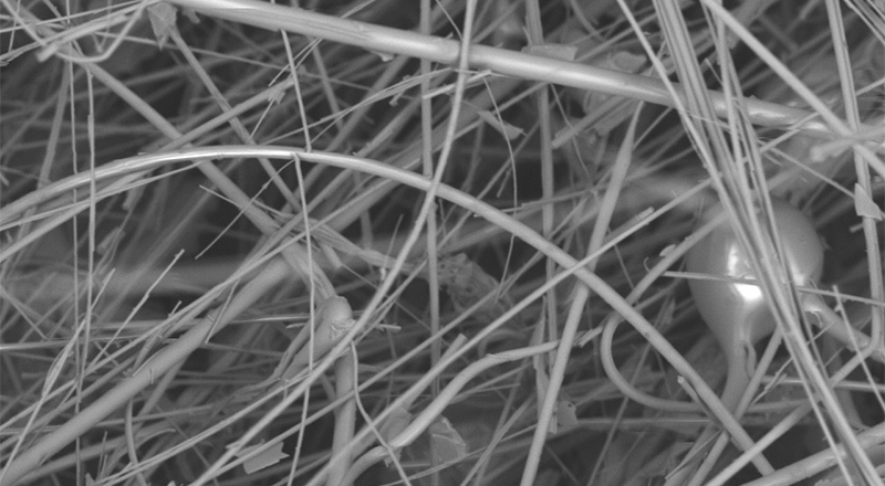
Current manufacturing involves a flow of molten minerals (at ca 1600°C) through which air is forced.  This creates fibres of amorphous structure that can be compressed together without binders.  More advanced production rapidly spins molten minerals similar to the production of candy floss, or cotton candy. This results in a mass of fine, intertwined fibres with a typical diameter of 2µm to 6µm (microns).


Credit: Knauf.com


High-Temperature Mineral Wool


High temperature mineral wools are rated for about 650°C to 1600°C and are made in similar ways to the lower temperature versions.  However, they are more expensive and so are used in refractory circumstances including kiln forming.

The three main types of HTIWs include:

Low Bio-persistent (LBP) Wool, including Alkaline Earth Silicate (AES) wools and others:

Alkaline earth silicate (AES) wool
       Calcium magnesium silicate wool
       Calcium silicate wool
       Magnesium silicate wool
Alkali metal silicate (AMS) wool
       Potassium alumino silicate wool

Alumino Silicate Wool (ASW), also known as Refractory Ceramic Fibres (RCF)
       Aluminium silicate wool
       Aluminium zirconium silicate wool

Polycrystalline Wool (PCW)
       Aluminium oxide wool
       Mullite wool

The main forms that kilnformers are interested in are blanket, paper and board.  The paper and board normally contain binders ranging from latex to cellulose. There are other forms: bulk fibres, modules or blocks formed ready for installation, vacuum formed shapes, cement mastics, textiles, yarns and ropes.


A brief description of these kinds of refractory mineral wools are:

Alkaline earth silicate wool (AES)

AES wool consists of amorphous glass fibres that are produced by melting a combination of calcium, magnesium oxides and silicone dioxide.  Products made from AES are generally used in equipment that continuously operates and in domestic appliances. AES wool has the advantage of being bio-soluble—it dissolves in bodily fluids within a few weeks and is quickly cleared from the lungs and so has been excluded from carcinogenic classifications. It is generally rated up to 1200°C.

Alumino silicate wool (ASW)

This is also known as refractory ceramic fibre (RCF), again consisting of amorphous fibres produced by melting minerals and blowing air across the flow.  In this case, a combination of aluminium oxide and silicon dioxide.  It has a low thermal conductivity, and good resistance to chemicals. Alumino silicate wool is generally used at temperatures from 600°C to 1300°C  for intermittent operation, making it good for kilnforming. 

This was classified in Europe as a carcinogen category 2 – “Substances that should be regarded as if they are carcinogenic to humans” under the Dangerous Substances Directive in 1997. This was translated under CLP Regulation into a carcinogen category 1B “Known or presumed human carcinogen; presumed to have carcinogenic potential for humans, classification is largely based on animal evidence”.

Some of the trade names used are:
  • Kaowool®, a high-temperature mineral wool made from kaolin. It was one of the first types of high-temperature mineral wool and continues to be used. It can withstand temperatures to 1250°C. 
  • Cerablanket®, is a spun blanket manufactured from a high purity blend of alumina-silica and is classified up to 1315°C.
  • Cerachem® and Cerachrome® provide chemical stability and strength and have acoustic as well as thermal insulation characteristics. They are classified to 1426°C.

There are bio-soluble fibres produced under trade names such as Superwool® with temperature ratings of 1300°C and 1450°C.  Superwool® fibres are exonerated from carcinogen classification within Europe and not classified as hazardous by IARC or under any national regulations throughout the world.

Polycrystalline wool (PCW)

Polycrystalline wool was commercialised in the 1970’s and consists of fibres that contain more than 70% aluminum oxide. It is produced by sol–gel method from aqueous spinning solutions. The water-soluble green fibres obtained as a precursor are crystallized by means of heat treatment. This is produced in small quantities for specialised applications.  Its characteristics are that the fibres are of regular defined dimensions, it is chemically and thermally stable, with low shrinkage and high tensile strength, all with less dust produced in handling.  It is a more expensive process than producing RCW papers and blankets.

The polycrystalline wool is generally used at temperatures above 1300°C.  One trade name is Denka Alcen with a temperature rating up to 1600°C. Denka blankets are more resistant to acid and alkaline solutions than conventional alumino-silicate fibre blankets and have good thermal insulation characteristics.

Other than kilnforming, applications are in the ceramics, metals, petrochemicals, aerospace and automotive industry sectors. Typical PCW applications include use as support mats in catalytic converters and diesel particulate filters to reduce exhaust emissions, and as insulation in industrial high temperature furnaces for energy conservation, particularly in high temperature and/or chemically aggressive environments.

Credit: Alibaba.com


Kilnforming Refractory Papers

There are two fibre papers widely used in kilnforming: Papyros and Thinfire.  These are special cases of the RCF papers and deserve particular attention, although they are subsets of the previously described RCF wools.

Papyros
This is a fibre paper similar in thickness to cartridge paper.  It consists of  aluminium hydroxide, hydrated magnesium silicate (hazard classification: irritant), alumina borosilicate glass (hazard classification: irritant), wood pulp and resin (both binders).  None of the materials used in the composition of Papyros are classified as a possible carcinogenic substance.  It is recommended that eye, breathing and skin protection be used when handling the fired residue to reduce any irritation.  Washing after handling the dusts is recommended.


Thinfire
This fibre paper is also like cartridge paper in thickness and has a slightly finer texture than Papyros.  Its constituents are aluminium hydroxide, glass fibre, polyvinyl alcohol, cellulose, and polyamide resin.  Only the glass fibre is classified as an irritant.  The dust can be an irritant to eyes and skin.  If either are irritated, wash with large amounts of water. It is sensible to use breathing protection while handling the fired residue.


The materials used place both these fibre papers in the AES group of refractory fibres, which are biosoluble.  The use of hydrated magnesium silicate in Papyros gives an extremely small increased health risk over Thinfire.

Credit: cdc.com

Fibre Paper – Health and Safety

Mineral wool fibres and refractory ceramic fibres have been  classified as "possibly carcinogenic to humans" (Group 2B).  In contrast, the more commonly used vitreous fibre wools produced since 2000, including insulation glass wool, stone wool, and slag wool, are considered "not classifiable as to carcinogenicity in humans" (Group 3). The International Agency for Research on Cancer (IARC) elected not to make an overall evaluation of the newly developed fibres designed to be less bio-persistent such as the alkaline earth silicate (AES) or high-alumina, low-silica (ASW) wools. 


Bio-soluble fibres are produced that do not cause damage to the human cell. These newer materials have been tested for carcinogenicity and most are found to be non-carcinogenic.

Due to the mechanical effect of fibres, mineral wool products may cause temporary skin itching. To diminish this and to avoid unnecessary exposure to mineral wool dust, information on good practices is available on the packaging of mineral wool products with pictograms or sentences. Safe Use Instruction Sheets like safety data sheets are also available from each producer.

People can be exposed to mineral wool fibres in the workplace by breathing them in, skin contact, and eye contact. … The National Institute for Occupational Safety and Health (NIOSH) has set a recommended exposure limit (REL) of 5mg/m3 total exposure and 3 fibres per cm3 over an 8-hour workday [the highest existing standard].  The equivalent European standard is set by the Registration, Evaluation, Authorisation and Restriction of Chemicals (REACH).

AES, ASW and PCW have been registered before the first EC deadline of 1 December 2010 and can, therefore, be used on the European market.
ASW/RCF is classified as carcinogen category 1B.
AES is exempted from carcinogen classification based on short-term in vitro study result.
PCW wools are not classified; self-classification led to the conclusion that PCW are not hazardous.

Based on the total experience with humans and the findings of scientific research (animals, cells), it can be concluded that elongated dust particles of every type have in principle the potential to cause the development of tumours providing they are sufficiently long, thin and bio-persistent. According to scientific findings inorganic fibre dust particles with a length-to-diameter ratio exceeding 3:1, a length longer than 5μm (0.005 mm) and a diameter smaller than 3μm (WHO-Fibres) are considered health-critical.

High-temperature mineral wool is processed into products containing fibres with different diameters and lengths. During handling of high-temperature mineral wool products, fibrous dusts can be emitted. These can include fibres complying with the WHO definition.

There is concern about the silica content of refractory fibres.  The silica that is of concern is of a crystalline structure.  The method of production does not produce crystalline silica. The process used to create the fibres is:
Amorphous high-temperature mineral wool [fibres] (AES and ASW) are produced from a molten glass [or mineral] stream which is aerosolised by a jet of high-pressure air or by letting the stream impinge onto spinning wheels. The droplets are drawn into fibres; the mass of both fibres and remaining droplets cool very rapidly so that no crystalline phases may form.

The potential effects on health of the materials in refractory fibres have been tested and found to be non-hazardous.

In after-use high-temperature mineral wool crystalline silica crystals are embedded in a matrix composed of other crystals and glasses. Experimental results on the biological activity of after-use high-temperature mineral wool have not demonstrated any hazardous activity that could be related to any form of silica they may contain.

Thus, no crystalline silica is produced and the risk of silicosis from refractory fibres does not exist.  Certain sizes of any fibre present other risks.

Risks


Consideration of risks and therefore precautions, relate to three factors: Dimension, Durability and Dose.

Dimension

Fiber dimensions are critical, as only fibres of a certain size can reach the lungs…. Mineral fibres with a diameter greater than 3 microns are, in humans, “non respirable”. … Even below this respirability threshold only the finest fibres may be deposited into the gas exchange region of the lungs.

While respirability is determined by fiber diameter, fiber length is also important. Short fibres behave as if they are compact particles and can be cleared by the normal mechanisms which involve cells called macrophages. However long fibres [greater than 5 microns] frustrate this mechanism and, for some still unknown reason, are more biologically active.

Durability

Durability in this context describes the ability of a material to persist in the body and so is more accurately called “bio-persistence”. …  Fibres can dissolve or they may break into shorter pieces which can then be removed to the airways or through the lymphatic system. The rate of removal of different fibres is typically measured … and expressed as their “half-life” – that is the time it takes to reduce the number of fibres in the lungs by 50%.

Dose

The [dose] is the result of [dimension and durability] and is often referred to as “lung burden”.  With chronic exposures the lung burden is the result of … [continued exposure] and … bio-persistence. If the exposure is high enough and clearance slow then a sufficiently large dose will accumulate for adverse health effects to result.


The scientific knowledge about fiber toxicity allows comparison of fibres in terms of their toxicological potency and has also driven several initiatives to reduce potential risks in the workplace.  This has led to development of manufacturing processes for thicker fibres, although this is limited by the lesser thermal efficiency of thick fibres.  Thicker fibres are also more likely to cause skin irritation.  A lot of effort has been put into the development of bio-soluble fibres such as the AES wools which are increasingly available.

Recent research has shown a gradation of increasing bio-persistence is in the order of – least to greater –
AES (Calcium Silicate);
AES (Magnesium Silicate);
PCW;
RCF. 
This same research shows that fibres longer than 20 microns cannot be easily cleared from the lungs.  Breathing protection must filter out all particles larger than 20 microns. 

The WHO research shows that lung health effects can be produced by particles down to 3 microns. This means that filters used must be able to eliminate particles larger than 3 microns to provide effective protection against high exposure.


Handling practices

Sensible precautions when handling refractory fibre papers are eye, breathing and skin protection.  This can be safety goggles, dust mask (see filter size above), and long gloves and long sleeves.  Higher levels of protection can be used, but are not indicated as necessary by the research and classifications of health and safety organisations in the western world.

During clean-up the fibres should be dampened before any brushing of the residue, or vacuumed with HEPA filters to reduce the movement of fibres into the air.  You should also wash exposed skin after handling any of the dust.  Clothes should also be cleaned and washed frequently. 

Do not smoke, eat or drink in areas where the fibre dust is present.


More detailed information is available in the e-book: Low Temperature Kilnforming.

The understanding of the composition and manufacture of refractory fibre papers and blankets should help assess the small risks of using these materials, and the precautions that should be taken in handling both the un-fired and fired forms.