Showing posts with label Medicine. Show all posts

Wednesday, 12 November 2025

Glass Microspheres in Medicine


Using Embolic Glass Microspheres to Target Chronic Disease

By Sierra Kucko


Glass Microspheres: The Tiny Superheroes of Glass Form Factors

Two main factors contribute to the properties of glass: composition and form factor. While emphasis is often placed on glass composition, the form factor is arguably equal in importance. Glass in the form of microspheres has permeated various industries, ranging from aerospace to medical sectors. MO SCI specializes in the production of precision glass microspheres that have become invaluable to these industries, and their usage is ever-evolving. Whether it be controlling gaps for adhesive bondline spacing, improving the visibility of road markings, or drug delivery devices, glass microspheres fit the bill.

Targeted Treatment with Embolic Glass Microspheres

Biocompatible (and in some cases biodegradable) microspheres are especially appreciated for medical applications, such as transarterial embolization (TEA) or musculoskeletal (MSK) embolization.1,2

TEA refers to the blockage of blood supply, which may sound like a bad thing, but in many cases, these are lifesaving procedures. For example, a substantial driver for this technology is cancer treatment. One way to combat a tumor or abnormal tissue growth is to cut off its blood supply, which can be achieved through the precise application of appropriately sized microspheres to occlude the fine vasculature ‘feeding’ it.1

Similarly, MSK embolic microspheres are sought after to prevent the abnormal overgrowth of blood vessels, a consequence of chronic inflammation. This kind of inflammation is part of a pathological loop, whereby the inflammation promotes the formation of new blood vessels that in turn, can feed nerve growth and contribute to chronic, debilitating pain.2 Microsphere embolization can therefore be used as a pain management tool, as well. For applications with this level of weightiness, the microsphere size is a chief feature.

Together with the form factor, glass microspheres can be tailored through their composition. First and foremost, any implantable glass must be compatible with the body. Ancillary to this, the composition can be altered to offer additional functionality. Using TAE as an example to put this concept into context, the composition of glass used in this type of application is unique and important.

TAE is a procedure utilized by interventional radiologists. Interventional radiology (IR) is the diagnosing and/or treatment of cancer and other conditions while avoiding major surgery. To achieve this, small tools such as needles, catheters, or wires are utilized in conjunction with radiation like MRI, ultrasound, etc. to apply treatment precisely to the tissue site.1–3 Personalization and optimization of outcomes is a clinical challenge of any medical intervention, making the accurate delivery and distribution of the embolic particles in real-time indispensable.

Due to the use of radiation to guide the placement, the embolic particle should be radiopaque (opaque to radiation) to ensure that guided delivery to the site can be realized. Compared with glass, this radiopacity is lacking or more difficult to achieve in microspheres derived from other material types.

Partner with MO SCI for Precision Glass Microspheres

Each application of glass is unique and therefore may require unique chemistries and form factors. Glass microspheres are becoming increasingly popular, since their form factor alone may improve the function of the glass (depending on the application) when compared to their powder or frit counterparts.

For applications that require precise microsphere size and composition, it is important to turn to trusted experts. MO SCI produces a wide range of glass microspheres in a variety of chemistries to suit nearly any need. Contact us today to learn how glass microspheres may be beneficial for your application.

References

  1. Pérez-López A., et al. (2022). Embolization therapy with microspheres for the treatment of liver cancer: state-of-the-art of clinical translation. Acta Biomaterialiahttps://doi.org/10.1016/j.actbio.2022.07.019

  2. Gremen E., et al. (2022). Safety and efficacy of embolization with microspheres in chronic refractory inflammatory shoulder pain: a pilot monocentric study on 15 patients. Biomedicines. https://doi.org/10.3390/biomedicines10040744

  3. Kishore S, et al, (2021). Transarterial embolization for the treatment of chronic musculoskeletal pain: a systematic review of indications, safety and efficacy. ACR Open Rheumatology. https://doi.org/10.1002/acr2.11383


Sunday, 15 May 2022

Using Silver-Releasing Glass to Reduce Bioburden


Using Silver-Releasing Glass to Reduce Bioburden

Posted  on 


Silver has been used for wound management for over 200 years, and its anti-microbial properties have been known since the 19th century.1 Although its direct use in the treatment of wounds fell out of favor when antibiotics were introduced, it continued to see applications in treatment for burns and other conditions on the surface of the body.

Recently, interest in silver has been renewed following research involving the use of silver with bioactive glasses that are implanted directly into the body. The unique properties of the glass and silver material allow it to be deployed in areas where antibiotics cannot reach; while the glass containment also allows more control over the concentration of silver ion released in specific areas.

Challenges with antibiotic resistance

A serious problem in all countries around the world is the rise of antibiotic-resistant bacteria that can cause issues for patients before, during, and after surgery. This includes strains such as methicillin-resistant Staphylococcus aureus (MRSA), that form biofilms on hospital equipment and surgical implants, increasing the bioburden on these surfaces.2

This is a particular problem for surgical implants: medical implant failure is commonly caused by infections resulting from bacteria living on implant surfaces. Due to the position of the implants and nature of the bacteria, these biofilms can be difficult to eliminate using antibiotics alone and treatment may require surgical removal.

How does silver impact bacteria?

One method of inhibiting the growth of antibiotic-resistant strains of bacteria on surgical implants is to coat them with silver-releasing glass. These glasses have been shown to be effective in reducing bacterial adhesion at the surface of implants, and the addition of silver further inhibits the development of biofilms on implant surfaces.3

The exact mechanism of how silver impacts bacteria is debated among scientists, but it’s generally accepted that the antibacterial action involves the release of Ag+ ions that interact to disrupt pathogens, compromising their ability to successfully replicate.4,5

Producing silver releasing glass

Conventional melt-quenching methods have proved successful in producing silver-doped glasses, however, difficulties in producing controlled and reproducible levels of silver below the allowed tolerance in humans prevent these techniques from being widely adopted. Other methods such as a sol-gel route have been explored – this enables much finer control over the introduction of the silver into the material structure.6

The future of research into these materials involves ensuring that there is sufficient silver to provide effective protection, while preventing the silver from leaching too quickly into the body and causing separate issues.

One promising method of activation is the use of phosphate-based glasses, which are soluble materials that allow for the controlled delivery of the silver ions.5 By incorporating the ions into the structure of the glass the two become a single phase and the rate of release of the silver is determined by the speed at which the glass degrades.

Phosphate-based glasses have already proven to be effective in delivering silver ions to help control urinary tract infections in patients with long-term indwelling catheters, as well as being used in wound dressings to prevent infections.5

The number of people requiring implant surgery is set to increase as life expectancy of the world’s population is expected to increase. This makes research and development of silver releasing glasses all the more important, and the procurement of high-quality research materials is vital.

Mo-Sci has extensive experience in the manufacture of biomedical glasses for healthcare, with a number of options for direct purchase. These biomedical glasses are available in sizes ranging from a few microns up to millimeter-sized structures depending on the form of the glass, and can be made into a range of shapes including microspheres, porous structures, and powders.

Custom solutions are also produced with Mo-Sci’s expert team of engineers and technicians to research, develop and produce glass which is custom-made to fit a wide range of applications. Contact us for more information.

References

  1. Clement, J. L. & Jarrett, P. S. Antibacterial Silver. Met. Based. Drugs 1, 467–482 (2007). https://www.ncbi.nlm.nih.gov/pmc/articles/PMC2364932/
  2. Valappil, S. P., Knowles, J. C. & Wilson, M. Effect of Silver-Doped Phosphate-Based Glasses on Bacterial Biofilm Growth. 74, 5228–5230 (2008). https://aem.asm.org/content/74/16/5228
  3. Cabal, B. et al. A new biocompatible and antibacterial phosphate free glass-ceramic for medical applications. Sci. Rep. 4, 1–9 (2014). https://www.nature.com/articles/srep05440
  4. Agostino, A. D. et al. Seed mediated growth of silver nanoplates on glass: exploiting the bimodal antibacterial effect by near IR photo-thermal action and Ag + release †. 70414–70423 (2016). https://pubs.rsc.org/en/content/articlehtml/2016/ra/c6ra11608f
  5. Valappil, S. P. et al. Effect of Silver Content on the Structure and Antibacterial Activity of Silver-Doped Phosphate-Based Glasses. 51, 4453–4461 (2007). https://www.ncbi.nlm.nih.gov/pmc/articles/PMC2168012/
  6. Rahaman, M. N. Bioactive ceramics and glasses for tissue engineering. Tissue Engineering Using Ceramics and Polymers: Second Edition (2014). doi:10.1533/9780857097163.1.67 https://www.sciencedirect.com/science/article/pii/B978085709712550003X
  7. Mo-Sci Glass Products https://mo-sci.com/en/products

Sunday, 6 March 2022

Reducing Implant-Related Infection with Bioactive Glass


Reducing Implant-Related Infection with Bioactive Glass

Posted  on 


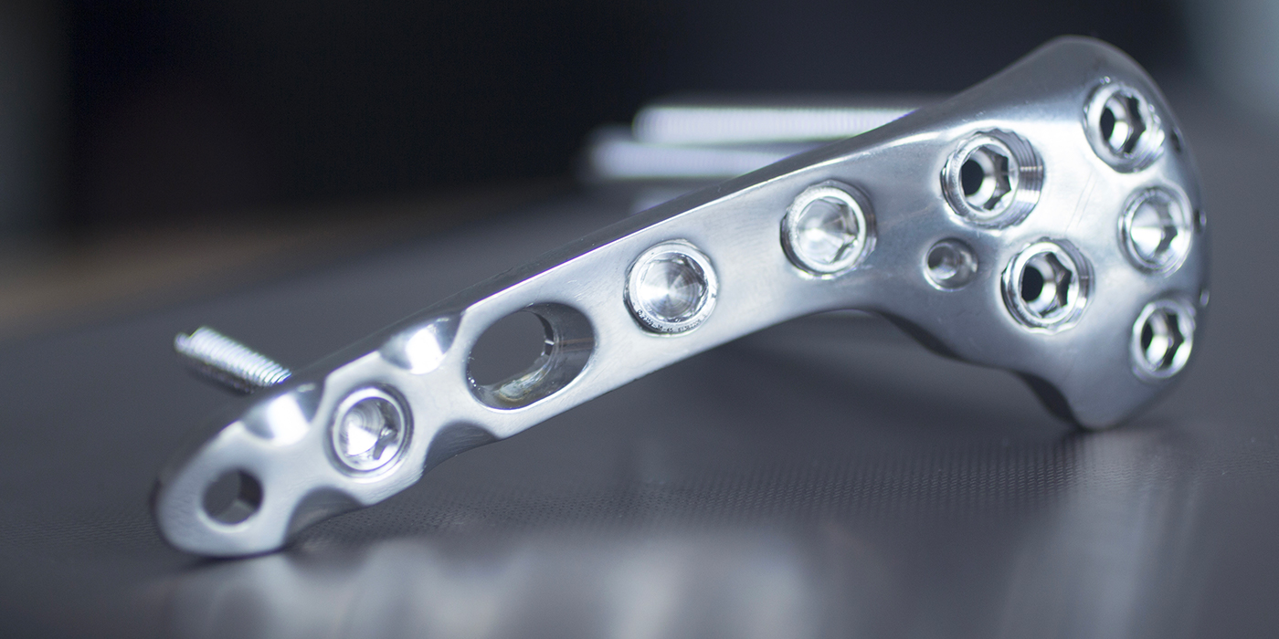
Close-up photo of an orthopedic implant

Disease, trauma or serious infection may result in extensive bone damage or bone loss which exceeds the body’s capacity to repair itself. In such cases, implants are needed to promote satisfactory healing. These may take the form of screws, plates, or rods to immobilize broken bones in the correct alignment, reinforce weak bones or correct skeletal deformities. Similarly, diseased joints can be replaced with prosthetic joints to restore normal, pain-free movement. 

Despite ongoing medical advances and improvements in materials and procedures, there remains a substantial risk of implanted devices becoming infected. In addition to microbes being introduced into the body during surgery, there is the risk of bacteria transported in the blood from other parts of the body colonizing the surface of an implant. It has been estimated that as many as 2.5% of primary hip and knee replacements and to 10% of joint revision surgeries are complicated by infection.1 Infected implanted devices represent a significant clinical challenge. Typically, despite lengthy antibiotic treatments, it is often necessary for the infected implant to be surgically removed. This not only increases patient morbidity and dissatisfaction, but is also associated with substantial cost.2 

Antibiotics continue to be the mainstay strategy for both the prevention and treatment of implant infections. However, the power of antibiotics in the fight against infection is diminishing as many strains of potent bacteria are developing resistance to even the strongest antibiotics. Consequently, the risk of implanted devices becoming infected is on the rise and researchers are investigating novel ways to reduce such infections.

One strategy is based on the discovery that the majority of bacteria live in surface-bound microbial communities, rather than as free-swimming entities. On binding to the surface, bacteria secrete adhesion proteins that provide an irreversible attachment.3 Such bacterial biofilms account for over 80% of clinical microbial infections.2 It was therefore proposed that making the surface of implants unsuitable for bacterial colonization would dramatically lower infection rates. This can be achieved by coating the implant with bioactive glass.

The Antimicrobial Properties of Bioactive Glass

Bioactive glass is a type of glass made from high-purity chemicals, such as silica, calcium, and boron, which induce specific biological activity.4 Bioactive glass, by virtue of its high strength, low weight and biocompatibility, has been widely used in a range of biomedical applications, including tissue engineering, bone grafting, dental reconstruction and wound healing.5 

Such clinical experience has shown that borate bioactive glass possesses antimicrobial properties against a wide range of bacteria, including MRSA and E-coli.6,7 The antimicrobial efficacy is achieved though an increase in pH of the surrounding body fluids (which is stressful for bacteria) and because any bacteria that do approach are unable to adhere to bioactive glass and so cannot create microfilms on its surface.8,9 In vitro studies have confirmed that bioactive glass has strong anti-staphylococcal and anti-streptococcal activity.10,11

Since the antimicrobial action of bioactive glass arises from it creating an environment that is hostile to bacteria rather than requiring direct contact with the invading microbe in order to kill it, it is effective across a wide range of bacteria. Furthermore, the bacteria cannot adapt to such effects, and so no bacteria have been found to develop resistance to the antimicrobial effects of bioactive glass.9 

Coating Implants with Bioactive Glass

Initial technical challenges have been overcome and a range of titanium implants have been successfully coated with bioactive glass.12 Clinical use of implants coated with bioactive glass has given promising results in both orthopedic and dental applications. There was no evidence of the coated implants causing any adverse effects or inflammatory response in the surrounding tissue. Furthermore, the implants coated with bioactive glass were found to accelerate cell attachment and mineralization of the extracellular matrix, promoting more rapid bone growth. In addition, the proportion of bone-to-implant contact was significantly greater for implants coated with bioactive glass compared with traditional implants.13-15

Enhancing the Antimicrobial Efficacy of Bioactive Glass

Bioactive glass has good antimicrobial action, being effective against a broad spectrum of aerobic and anaerobic bacteria.9 However, the antimicrobial effects can be further enhanced to increase the range of antimicrobial activity, by the addition of ions, such as boron, copper, silver, yttrium, and iodine, or organic nanoparticles.16,17 The chosen antimicrobial agent is incorporated into the bioactive glass during its production and released once the bioactive glass is in an aqueous solution, creating an environment inhospitable for microbial life. The bioactive glass can thus be used as a delivery system for antimicrobials.18

The advantage of ions and nanoparticles over antibiotics is that their efficacy depends solely on contact with the bacterial cell wall; they do not need to enter the cell. Consequently, their lethal effect is delivered irrespective of the specific genetics of the target bacteria and is unaffected by the resistance mechanisms used by bacteria to evade antibiotics.

Conclusion

Infection of medical implants is an increasingly serious clinical and socioeconomic burden. Furthermore, the situation is likely to worsen with the increasing prevalence of bacteria with multi-drug resistance. 

Bioactive glass has inherent antimicrobial activity and does not elicit a toxic response to surrounding tissues. Consequently, coating implants with bioactive glass represents an attractive option for reducing the risk of infection. The antimicrobial properties of bioactive glass can be further enhanced by loading it with antimicrobial agents, such as ions or antibacterial nanoparticles. Such a strategy would reduce the need for prophylactic antibiotic use, whereby protecting against the development of further strains of antibiotic-resistant bacteria. 

Since bacteria cannot adapt to the hostile environment created by bioactive glass or its biofilm-resistant surface, they are unlikely to develop resistance to the antimicrobial action of bioactive glass. 

In addition, the coating of implants with bioactive glass has been shown to speed up the fusion of the implant with bone, accelerating a patient’s recovery.

The use of bioactive glass, either alone or doped with antimicrobial agents, as a coating for orthopedic and dental implants is thus likely to improve the success rate and enhance patient outcomes across a range of reparative and restorative surgeries by promoting rapid healing and minimizing the occurrence of infection. 

Mo-Sci produces high quality bioactive glass powders, the precise composition of which can be tailored to meet specific requirements. They produce bioactive glass suitable for coating orthopedic and dental implants.

References & Further Reading

  1. Lentino JR. Prosthetic joint infections: Bane of orthopedists, challenge for infectious disease specialists. Clin Infect Dis. 2003;36:1157–61. doi: 10.1086/374554.
  2. Hall-Stoodley L, et al. Bacterial biofilms: from the natural environment to infectious diseases. Nature Reviews Microbiology2004;2(2): 95–108.
  3. Davey ME and O’Toole GA. Microbial biofilms: from ecology to molecular genetics. Microbiology and Molecular Biology Reviews2000;64(4):847–867.
  4. Brauer DS. Bioactive Glasses—Structure and Properties. Angew Chem Int Ed 2015;54: 4160–4181.
  5. Rahaman MN, et al. Bioactive glass in tissue engineering. Acta Biomaterialia 2011;7:2355?2373.
  6. Ottomeyer M, et al. Broad-Spectrum Antibacterial Characteristics of Four Novel Borate-Based Bioactive Glasses. Advances in Microbiology 2016;6:776?787.
  7. Khvostenko D, et al. Bioactive glass fillers reduce bacterial penetration into marginal gaps for composite restorations. Dental materials 2016;32(1):73–81. Available at http://www.demajournal.com/article/S0109-5641(15)00437-6/pdf
  8. Zhang D, et al. Factors Controlling Antibacterial Properties of Bioactive Glasses. Key Engineering Materials 2007;330-332:173?176.
  9. Drago L, et al. Recent Evidence on Bioactive Glass Antimicrobial and Antibiofilm Activity: A Mini-Review Materials 2018;11:326?337.
  10. Misra SK, et al. Poly(3-hydroxybutyrate) multifunctional composite scaffolds for tissue engineering applications. Biomaterials 2010;31:2806–2815.
  11. Rivadeneira J, et al. In vitro antistaphylococcal effects of a novel 45S5 bioglass/agar-gelatin biocomposite films. J Appl Microbiol 2013;115,604–612.
  12. Lopez-Esteban S, et al. Bioactive glass coatings for orthopedic metallic implants. Journal of the European Ceramic Society 2003;23:2921–2930.
  13. Mehdikhani-Nahrkhalaji M, et al. Biodegradable nanocomposite coatings accelerate bone healing: In vivo evaluation. Dent Res J (Isfahan). 2015;12(1):89?99.
  14. Chen Q, et al.Cellulose Nanocrystals–Bioactive Glass Hybrid Coating as Bone Substitutes by Electrophoretic Co-deposition: In Situ Control of Mineralization of Bioactive Glass and Enhancement of Osteoblastic Performance. ACS Appl Mater Interfaces. 2015 Nov 11;7(44):24715?25.
  15. van Oirschot BA, et al. Comparison of different surface modifications for titanium implants installed into the goat iliac crest. Clin Oral Implants Res. 2016;27(2):e57?67.
  16. Kaur G, et al D. Review and the state of the art: Sol–gel and melt quenched bioactive glasses for tissue engineering. J Biomed Mater Res B Appl Biomater 2016;104, 1248–1275.
  17. Karwowska E. Antibacterial potential of nanocomposite-based materials – a short review. Nanotechnology Reviews 2016;6(2):243?254.
  18. Rivadeneira J and Gorustovich J. Bioactive glasses as delivery systems for antimicrobial agents. Journal of Applied Microbiology 2016;122, 1424–1437.
  19. Mo Sci Corporation website. http://www.mo-sci.com/en/products

Sunday, 27 February 2022

Inhibiting Bacterial Growth with Bioactive Glass


Inhibiting Bacterial Growth with Bioactive Glass

Posted  on 


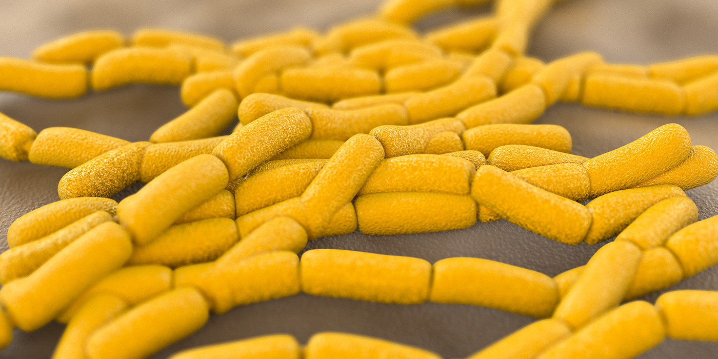
Computer rendering of bacterial culture

Despite numerous advances in technologies, bacteria remain a cause for concern in both clinical and industrial settings. They pose an especially challenging problem in medicine where only bactericidal strategies which have a minimal detriment to human health are feasible, and bacterial infection continues to be a leading cause of morbidity and mortality.

Surgical procedures in particular are associated with an increased risk of infection, and infection is the most frequently encountered post-surgical complication. The primary means of prevention and treatment of bacterial infections is the administration of antibiotics. However, the power of antibiotics in the fight against infection is diminishing as strains of potent bacteria are developing multi-drug-resistance.

In contrast to historical beliefs, it is now apparent that the majority of bacteria live in surface-bound microbial communities, rather than as free-swimming entities. On binding to the surface, bacteria secrete adhesion proteins that provide an irreversible attachment. The bacteria proceed to proliferate and create a colony that ultimately results in the formation of a mature biofilm protected by a peptidoglycan envelope.1 Such biofilms account for over 80% of clinical microbial infections, and so are associated with considerable morbidity and expense.2

In addition to the possibility of biofilms containing antibiotic-resistant strains, the bacteria are protected from antibacterial agents and the body’s immune system by the peptidoglycan envelope. Biofilm-associated bacteria are 100 to 1,000 times less susceptible to antibiotics than free-swimming bacteria, and so patients with biofilm infections are rarely cured by treatment with antibiotic agents, which are used nonetheless due to the lack of alternatives. Furthermore, there is the scope for bacteria to leave the biofilm and colonize other areas throughout the body.

Consequently, there has been much research in to the development of new broad-spectrum antibiotics and also novel antimicrobial strategies.

Novel antimicrobial materials

Nanotechnology has opened up the potential for the development of new types of materials with antimicrobial properties. It allows the physicochemical properties of a material to be changed in order to achieve antimicrobial effects.3

Antimicrobial nanomaterials include a wide range of metal, metal oxide, and organic nanoparticles and so have numerous modes of action. Although a range of chemical interactions are involved, the end result is membrane damage that leads to loss of integrity and impaired metabolism and ultimately cell death.

The advantage of nanoparticles over antibiotics is that their efficacy depends solely on contact with the bacterial cell wall; they do not need to enter the cell. Consequently, their lethal effect is exerted irrespective of the specific genetics of the target bacteria and is unaffected by the resistance mechanisms used by bacteria to evade antibiotics. Furthermore, the large surface area to size ratio of nanoparticles means that high activity can be achieved with a small dose, whereby minimizing the risk of toxicity. It is therefore hoped that nanoparticles may provide an effective alternative to antibiotics for the treatment of both free-swimming and surface bound bacteria.

Nanoparticles could be used in antimicrobial treatments and in the manufacture of nanocomposite products suitable for use in medical materials and devices.

Bioactive glass

Bioactive glass is a type of glass made from high-purity chemicals, such as silicon oxide, calcium oxide, and phosphorus oxide, that induces specific biological activity.4 Furthermore, by modifying the composition and structure of the glass, its physical properties can be tailored to meet a specific need.4

Bioactive glass elicits a negatively benign immune response and has been widely used in a range of biomedical applications, including tissue engineering, bone grafting, dental reconstruction and wound healing.5 It is able to bond to either hard or soft tissue and has been shown to facilitate strong new bone growth, promote soft tissue regeneration and enhance vascularization to ensure a healthy blood flow to the newly regenerated tissue.6–8 It has also been mixed with dental filling materials to promote the remineralization of dental caries.9

In addition, borate bioactive glass has been shown to have antimicrobial properties against a wide range of bacteria, including MRSA and E-coli.10 It has been shown that bacteria are unable to adhere to bioactive glass and so microfilms cannot develop on its surface.11 This is supported by clinical observations; no infections have been reported on bioactive glass implants.11 In addition, the inclusion of bioactive glass into dental filling material reduced bacterial penetration by 40%, whereby reducing the rate of decay and increasing the lifetime of the restoration.12

The antimicrobial action of bioactive glass can be further increased, giving the glass a broader spectrum of antimicrobial activity, by the addition of ions such as silver, yttrium, selenium, and iodine.13

The use of bioactive glass fibers to promote wound healing and soft tissue repair and the inclusion of bioactive glass in bone grafting and dental restoration composite materials to promote bone growth thus gives dual benefit — more rapid healing and reduced risk of infection.

Mo-Sci produces implant grade bioactive glass powders of varying sizes and with specific compositions suitable for mixing with composite materials.14

References

  1. Davey ME and O’Toole GA. Microbial biofilms: from ecology to molecular genetics. Microbiology and Molecular Biology Reviews 2000;64(4):847–867.
  2. Hall-Stoodley L, et al. Bacterial biofilms: from the natural environment to infectious diseases. Nature Reviews Microbiology 2004;2(2): 95–108.
  3. Karwowska E. Antibacterial potential of nanocomposite-based materials – a short review. Nanotechnology Reviews 2016;6(2):243?254.
  4. Brauer DS. Bioactive Glasses—Structure and Properties. Angew Chem Int Ed 2015;54: 4160–4181.
  5. Rahaman MN, et al. Bioactive glass in tissue engineering. Acta Biomaterialia 2011;7:2355?2373.
  6. Gerhardt L-C and Boccaccini AR. Bioactive Glass and Glass-Ceramic Scaffolds for Bone Tissue Engineering. Materials 2010;3:3867?3910
  7. Pugely AJ, et al. Influence of 45S5 Bioactive Glass in A Standard Calcium Phosphate Collagen Bone Graft Substitute on the Posterolateral Fusion of Rabbit Spine. Iowa Orthop J. 2017; 37: 193–198.
  8. Gorustovich A, et al. Effect of bioactive glasses on angiogenesis: In-vitro and in-vivo evidence: A review. Tissue Eng. Part B Rev. 2010;16:199?207.
  9. Chatzistavrou X, et al. Fabrication and characterization of bioactive and antibacterial composites for dental applications. Acta Biomater. 2014;10:3723–3732. Available at https://www.ncbi.nlm.nih.gov/pubmed/24050766
  10. Ottomeyer M, et al. Broad-Spectrum Antibacterial Characteristics of Four Novel Borate-Based Bioactive Glasses. Advances in Microbiology 2016;6:776?787.
  11. Zhang D, et al. Factors Controlling Antibacterial Properties of Bioactive Glasses. Key Engineering Materials 2007;330-332:173?176.
  12. Khvostenko D, et al. Bioactive glass fillers reduce bacterial penetration into marginal gaps for composite restorations. Dental materials 2016;32(1):73–81. Available at http://www.demajournal.com/article/S0109-5641(15)00437-6/pdf
  13. Xu Y, et al. Study on the Preparation and Properties of Silver-Doped Borosilicate Antibacterial Glass. Journal of Non-Crystalline Solids 2008;354:1342?1346.
  14. Mo Sci Corporation website. http://www.mo-sci.com/en/products.

Sunday, 13 February 2022

Using Porous Glass Microspheres for Targeted Drug Delivery


Using Porous Glass Microspheres for Targeted Drug Delivery

Posted  on 

linkedin sharing button Share
twitter sharing button Tweet
facebook sharing button Share

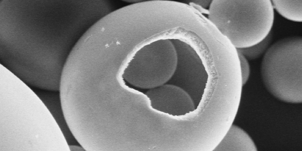
Porous hollow glass microspheres

Finding methods for improving efficiency and bioavailability is central to targeted drug delivery. While controlled release dosage medication forms are the norm, they can encounter many limitations. One disadvantage is the difficulty of locating and retaining the drug delivery system within the gastrointestinal (GI) tract due to gastric emptying variation.1 This can cause drug release that is insufficient for the patient or result in shorter residence time of the dosage in the stomach. 

To increase gastric retention and improve drug absorption, hollow microspheres have been developed and applied in the clinical setting for certain patients.2 Porous-wall hollow glass microspheres used in medicine are often produced from biopolymers, ceramics, bioactive glasses, and silicates. Hollow microspheres feature a 10 to 100 micron-diameter hollow cavity for the containment of certain substances.3

The most predominant mode for drug administration to the systemic circulation is the oral route. Some drugs have difficulty absorbing through the GI tract when using this route, prompting professionals to seek alternative methods for delivering pharmacologically active substances to the body.

The porous or hollow features of microspheres offer the ability to encapsulate fragile drugs and provides protection from biological compounds that may interfere with drug availability.4 These spherical, empty particles can remain in the gastric region for long periods of time and extend residence time of drugs in the GI tract.

Porosity offers improved loading efficiency and helps control the release of medications. Overall, hollow microspheres improve bioavailability of a drug, thereby reducing drug waste.

Microspheres can be produced to feature a uniform shape and size that can improve delivery of spheres to a specific target site. Additionally, microspheres are an ideal candidate for carriers of therapeutic agents due to their porosity, large surface area, and volume.

The hollow center of the microspheres reduces their density to such a degree that they have the potential to be buoyant. This behavior makes hollow microspheres suitable for use in a wide variety of applications.

Advantages of Porous Glass Microspheres

Hollow microspheres can reduce dosing frequency, allowing for improved patient compliance. Also, a desirable plasma concentration of a therapeutic agent can be maintained with microspheres by continuous drug release. The structure of the microspheres also results in an increase in gastric retention time, meaning therapeutic agents are released over a longer time. Other advantages of hollow microspheres include:

  • Site-specific drug delivery to the stomach
  • Sustained release effect helps avoid gastric irritation
  • Short half-life drugs can achieve a better therapeutic effect

Limitations of Porous Glass Microspheres

Patients are required to consume a large amount of water for hollow microspheres to float and function. Instead of merely taking a sip of water with a drug (typical of the oral route of medication administration), patients must drink a full glass (200 to 250 ml) of water with the microsphere. In addition, hollow microspheres are unsuitable for drugs that feature stability and solubility issues in gastric fluids.

Savannah River National Lab (SRNL) and Mo-Sci Microspheres

The SRNL has teamed with Mo-Sci Corporation, experts in the production of specialty glasses, to create porous wall, hollow glass microspheres that consists of glass “microballoons” smaller than the diameter of a human hair.

SRNL’s microspheres feature a unique network of interconnected pores in the walls. These pores allow tiny “microballoons” to be filled with gasses as well as other materials. Each SRNL microsphere is around 50 microns in diameter, and the walls are around 10,000 angstroms thick. The walls feature pores that come to about 100 to 300 angstroms. Each pore allows gasses to entire the spheres and be stored or cycled on absorbents. 

The original use for SRNL’s microspheres was to provide a solid-state storage method for hydrogen. Further research, however, has revealed their powerful utility and practical application in medicine. Currently, Mo-Sci plans to offer SRNL’s porous-walled hollow glass microspheres as a transport system for targeted drug delivery, among other uses. 

Microspheres are being further investigated in an effort to discover new applications within healthcare. While further study is ongoing, microspheres will hopefully provide enhanced patient care and improve the effectiveness of medication delivery across a broad range of disease states.

References

  1. Kurrey A, Suresh PK, Singh MR. Hollow microspheres as a drug carrier: An overview of fabrication and in vivo characterization techniques. Chronicles of Young Scientists. 2014;5(1):1-10.
  2. Qing W, Wenhai H, Deping W. Preparation of hollow porous HAP microspheres as drug delivery vehicles. Journal of Wuhan University of Technology-Mater. Sci. 2007;22(1):174177.
  3. Hossain KMZ, Patel U, Ahmed I. Development of microspheres for biomedical applications: a review. Progress in Biomaterials. 2015;4(1):1-19.
  4. Li S, Nguyen L, Xiong H. Porous-wall hollow glass microspheres as novel potential nanocarriers for biomedical applications. Nanomedicine. 2010;6(1):127–136.
  5. Wicks GG, Heung LK and Schumacher RF. Microspheres and Microworlds. American Ceramic Society Bulletin, Vol. 87, No. 6
  6. http://www.mo-sci.com/porous-silica/ 

Sunday, 6 February 2022

Bioactive Glass as a Bone Graft Substitute


Bioactive Glass as a Bone Graft Substitute

Posted  on 


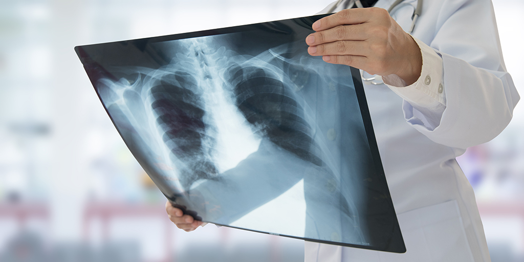
Doctor holding x-ray

Between 1992 and 2007, bone grafting was used in the treatment of almost two million patients in the United States.1 Bone grafts are used to facilitate the healing of complex bone trauma. This may be a multiple fracture or non-union of a long bone fracture, loss of bone due to disease, or surgical implantation of devices, like joint replacements, plates, or screws.

Bone grafting typically uses bone from another part of the patient’s body, such as ribs, hips, or pelvis, (an autograft) or bone harvested from a deceased donor or a cadaver that has been cleaned and stored in a tissue bank (an allograft). 

Despite grafting markedly improving bone regeneration and clinical outcomes after severe bone injury or loss, there is still room for improvement. Researchers continue to investigate ways to enhance bone grafting techniques and provide faster and denser bone regeneration with lower morbidity.

One such development has been the use of bone graft substitutes, such as demineralized bone matrix, calcium phosphates, collagen- /hydroxyapatite-based substitutes, and bone morphogenetic proteins. Although autologous bone grafting was considered to be the preferred bone grafting modality,2 there is a trend towards favoring artificial bone grafts over autografts since they are readily available and obviate the need for additional surgery. However, bone graft substitutes can have limitations regarding strength under torsion.

Bioactive glass, by virtue of its biocompatibility, strength and range of achievable properties, has been widely used to facilitate bone repair and provide support in tissue engineering.3

The Different Types of Bone Grafting

Bone grafting is a surgical procedure that is beneficial for repairing bones that have been severely damaged by trauma and for replacing bone that is missing as a result of either trauma or disease. It can also be used to strengthen bone at the site of an implant, be that a joint replacement, screw or dental implant. The bone graft provides a framework to support and encourage the growth of new, living bone.

Autografts have long been considered the preferred means of bone grafting since they do not carry the risk of rejection. However, these necessitate additional surgery, which increases patient morbidity and the risk of infection. Furthermore, there may be issues with availability finding a suitable site to harvest bone of the needed shape and size.

Allografts obviate the need for additional incisions but carry the risk of immune response preventing the graft from being accepted. There is also still the potential for availability issues since the donated bone needs to be tissue matched with the patient. 

A third option is not to rely on bone at all for the graft, but instead to use a man-made substitute. A range of different materials, including calcium phosphates, collagen, hydroxyapatite, have been investigated for use in bone grafting and are readily available. When using bone substitutes the nature of the materials must be carefully considered in terms of biocompatibility, resorption rate and strength.

Bone Graft Substitutes

The formation of new bone requires three key processes: osteogenesis (synthesis of new bone), osteoinduction (recruitment of stem cells and their differentiation into bone cells), and osteoconduction (the development of adequate blood supply to the new bone and correct structuring of the new bone cells). Bone graft substitutes are designed to facilitate and enhance these processes to promote rapid development of strong new bone.

Bone graft substitutes are frequently used to fill bone defects after orthopedic trauma. Ideally a synthetic bone graft substitute would have efficacy at least comparable to autograft, no immunogenicity, osteoinductive and osteoconductive properties, predictable resorption/degradation time, and no safety concerns. 

Numerous studies have reported benefits of using synthetic bone substitutes for fracture treatment and spinal surgery.4,5,6,7 These include reduced pain, bleeding and healing time, and improved functional outcomes compared with autografts. However, there have been safety concerns and problems with unpredictable resorption rates with some of the bone substitute materials.8

Furthermore, the different bone substitute products have varying characteristics. They all only provide minimal structural integrity and none targets all three of the key bone formation processes.9 Although some bone substitutes closely mimic the structure of natural bone, they lack osteogenic and osteoinductive properties. 

Bioactive glass has been successfully used in a range of tissue engineering procedures.3 With its versatility, achieved through the tailoring of properties through composition adjustments, its intrinsic strength and biocompatibility, bioactive glass was considered a prime candidate for improving synthetic bone substitutes.

Bioactive Glass for Bone Grafting

Introduction of bioactive glass into the body induces specific biological activity that causes soluble ionic species to be released. These lead to the glass becoming coated with a substance similar to hydroxyapatite. The formation of this layer allows bioactive glass to bond firmly with both hard and soft tissues. Furthermore, bioactive glass can be manufactured to release nutrients required for bone regeneration.

It has been shown that damaged bone regained its original strength more quickly when repaired using composite combined with bioactive glass compared with bone repair using composite alone and that the efficacy achieved is comparable to that of autologous bone grafting.10,11

A recent study compared spine fusion in rabbits using a mineralized collagen bone substitute with and without added bioactive glass. The bioactive glass-collagen composite was shown to closely mirror repair by autograft in terms of the amount and quality of the new bone.12 In addition, fusion occurred earlier when the collagen composite was augmented with bioactive glass.13

Conclusion

Bone grafting is an important tool for the repair of damaged or disease bone. The gold standard is autografting, which uses bone harvested from the patient to avoid rejection reactions. However, the increased morbidity caused by the additional surgery needed to acquire bone for grafting has resulted in an on-going quest to find an alternative. Bone substitutes have shown efficacy, but do not promote the formation of new bone. Bioactive glass is biocompatible and enhances strong new bone creation. Studies have now shown that the addition of bioactive glass to bone substitutes can increase their efficacy and bone healing characteristics to rival those achieved with autografting.

Mo-Sci produces medical implant grade bioactive glass in a form suitable for mixing with bone composites and can tailor its composition to meet specific requirements.13

References

  1. Kinaci A, et al. Trends in Bone Graft Use in the United States. Orthopedics 2014;37(9):e783 e788.
  2. Flierl MA, Outcomes and complication rates of different bone grafting modalities in long bone fracture nonunions: a retrospective cohort study in 182 patients. J Orthop Surg Res. 2013;8:33.
  3. Rahaman MN, et al. Bioactive glass in tissue engineering. Acta Biomaterialia 2011;7:2355 2373.
  4. Bajammal SS, et al. The use of calcium phosphate bone cement in fracture treatment: a meta-analysis of randomized trials. J Bone Joint Surg [Am] 2008;90-A:1186-1196.
  5. Swiontkowski MF, et al. Recombinant human bone morphogenetic protein-2 in open tibial fractures: a subgroup analysis of data combined from two prospective randomized studies. J Bone Joint Surg [Am] 2006;88-A:1258 1265.
  6. Lerner T, et al. A level-1 pilot study to evaluate of ultraporous beta-tricalcium phosphate as a graft extender in the posterior correction of adolescent idiopathic scoliosis. Eur Spine J 2009;18:170-9.
  7. Dimar JR, et al. Clinical and radiographic analysis of an optimized rhBMP-2 formulation as an autograft replacement in posterolateral lumbar spine arthrodesis. J Bone Joint Surg [Am] 2009;91-A:137 186.
  8. Carragee EJ, et al. A critical review of recombinant human bone morphogenetic protein-2 trials in spinal surgery: emerging safety concerns and lessons learned. Spine J 2011;11:471 191.
  9. American Academy of Orthopaedic Surgeons. Bone-grafts: Facts Fictions and Applications. Presented at 70th Annual General Meeting. Louisiana 2003. Available at https://www.aaos.org/research/committee/biologic/bi_se_2003-1.pdf
  10. Havener MB, et al. Improvements in Healing with a Bioactive Bone Graft Substitute in a Canine Metaphyseal Defect. Poster at 55th Annual Meeting of the Orthopaedic Research Society. February 22–25, 2009
  11. Jia W, et al. Bioactive Glass for Large Bone Repair. Adv Health Mater. 2015;4(18):2842 2848.
  12. Pugely AJ, et al. Influence of 45S5 Bioactive Glass in A Standard Calcium Phosphate Collagen Bone Graft Substitute on the Posterolateral Fusion of Rabbit Spine. Iowa Orthop J. 2017; 37: 193–198.
  13. Mo Sci Corporation website. http://www.mo-sci.com/en/products